• English
  • العربية
  • 中文
  • Français
  • Русский
  • Español

Dosimetry

Nuclear Medicine

Definition

The value of absorbed dose represents the radiation energy deposited in a tissue per unit mass. This quantity has units of grays (1 Gy = 1 J/kg). This value is very important in all medical procedures which use radiation because it serves as an indicator to predict tissue response. The dosimetry performed in nuclear medicine is called internal dosimetry. This name illustrates the fact that the doses are deposited by particles emitted from radionuclides located within the body. Hence, in order to properly calculate the absorbed dose to a tissue during a nuclear medicine procedure, we need to accurately assess the radionuclide distribution in the body, how many times those radionuclides decay, and how much of the energy they emit gets deposited in the tissue of interest.

The tissues where the radionuclides are located are called source tissues and the tissues where the energy is deposited are called target tissues. The amount of radionuclide that localizes in a source tissue is the uptake.   Source tissues can also be target tissues. Self-dose is the dose deposited in a source tissue by the radioactive decays occurring within it.  Source and target tissues are not implicitly a whole organ, they can be any region of interest as defined by the scientist (a group of organs, organ subregions, specific cell groups, or even cell organelles or molecules such as DNA).

Currently, the uncertainty associated with internal dosimetry calculations is very large compared to that of external therapy calculations. Expected errors in external beam doses are less than 5% while the most accurate internal dosimetry estimates one can currently achieve have errors of at least 20%.    

It is also important to note, that the term “dose” is used both in reference to the amount of activity administered to the patient and the absorbed dose imparted to a tissue.  The meaning of the word in each instance can be inferred by the context. The term “Effective dose” is also used in internal dosimetry documents. Effective dose and absorbed dose are calculated using the same procedure, only radiation weighting factors accounting for the differences between doses imparted by different types of particles and tissue weighting factors accounting for the radiation sensitivity of different organs are included in the effective dose calculations. For more information refer to the documents listed in the bibliography section.

General principles

Nuclear medicine is based around a fundamental concept of administering a radiopharmaceutical to a patient with the aim of achieving a diagnostic or therapeutic result. Like all medical procedures, the risk vs. benefit ratio needs to be understood, optimized and acceptable. What makes internal dosimetry more complicated than other types of radiation dosimetry calculations is the close proximity of the radioactivity source to organs and tissues, as well as the complexities involved in characterizing the source of the radioactivity and dosimetric volumes of interest, e.g. the activity distribution relative to the organs and tissues of interest.

There is no gold standard system for determining internal dosimetry, but there are several developed resources and methodologies available for estimating the dosimetric impact of clinical nuclear medicine practices.

Nuclear medicine dosimetry is used for clinical applications (both imaging and therapeutic procedures), radiation safety, and as well as for new drug development.

Important principles

The mean absorbed dose to an organ from a radiopharmaceutical is dependent on the characteristics of both the radionuclide and the pharmaceutical in terms of the type and amount of radiation emitted and the spatial and temporal distribution of the radiopharmaceutical in the body.The methodology most commonly used for internal dosimetry in humans in contemporary literature is the MIRD formalism, developed by the society of Nuclear Medicine  The other established methodology is that developed by the ICRP, which essentially is very similar to the approach used in the MIRD methodology. These approaches provide the methodology, terms, and equations necessary to determine radiodosimetry estimates, and can be used for situations where there is a known patient geometry.

The general equation for general absorbed dose calculations is the following:


For internal dosimetry, the numerator, E, is not simply derived because it depends on the activity and distribution of the radioactivity source relative to the volume of dosimetric interest and is thus not easily described.  For the sake of allowing manageable discrete calculations, sources and targets are usually summarized as source and target organs, or volumes or interest. In general E is calculated for every source affecting a target, and should account for 100% of relevant radioactivity. The value of E should incorporate:

  • Number of disintegrations from the source (usually summarized as source organs)
  • Energy of the disintegrations
  • Fraction of energy absorbed in target per disintegration of source (based on geometry, attenuation, and absorption properties)

Dose is often reported as calculated for particular/critical organs using absorbed dose (D) or equivalent dose (ED).  Overall dose to an individual can be reported as a total body dose or effective dose.  The MIRD pamphlet No. 21 provides an excellent clarification on the different values and nomenclatures which have been used in the field of radiopharmaceutical dosimetry, as well as an overview of the current state of nuclear medicine dosimetry. This article also encourages the nuclear medicine community to standardize the use of "equivalent dose" and "effective dose" for comparative evaluations of potential risks for nuclear medicine procedures.

Traditional dose calculation algorithms have calculated dosimetry for standard phantoms - human models with known geometries.  More recently, with the development of Monte Carlo and dose point kernel techniques, patient specific dosimetry is beginning to be studied and employed.

Because of the complexities involved in nuclear medicine dosimetry, as well as the array of methods and literature which has been presented, there are several organizations and web resources which provide overviews of the field - to a greater extent than what has been presented here.  Interested readers are advised to visit the following websites: the Medical Internal Radiation Dose (MIRD) Committee (SNM), the RAdiation Dose Assessment Resource (RADAR),  the Radiation protection of patients (RPoP -IAEA) - Nuclear Medicine section the International Commission on Radiological Protection (ICRP), and the National Council on Radiation Protection and Measurements (NCRP).

Patient dose optimization

For diagnostic nuclear medicine procedures, the patient exposure should be limited to the minimum necessary to achieve the clinical purpose of the procedure while providing acceptable image quality. For therapeutic nuclear medicine procedures, the appropriate radiopharmaceutical and activity are selected and administered so that the activity is primarily localized in the organs of interest, while the activity in the rest of the body is kept as low as achievable.

Important principles

For diagnostic procedures, it is necessary for the nuclear medicine specialist, in cooperation with the medical physicist, to determine the optimum activity to administer in a certain type of examination. In the case of nuclear medicine, DRLs are given as administered activity for a certain type of examination and for a normal size/age of patient. These guidelines assist in the optimization of protection by helping to avoid unnecessarily high activities for the patient or activities too low to provide useful diagnostic information.

In most countries, the weight  of the standard adult patient is assumed to be inthe range 70–80 kg. However, many patients fall outside of this range. If a fixed activity is used for all patients, this will lead to an unnecessarily high radiation exposure for a patient weighing less and images of unacceptable quality, or very long imaging times in obese patients.

There have been various approaches to determining the activity to be administered. These are usually designed to provide a constant count density in the image to maintain image quality or to provide a constant effective dose to the patient. For example, it has been shown that for myocardial perfusion scans using 99mTc-tetrofosmin, the activity should be increased by 150% for a 110 kg patient and by 200% for a 140 kg patient in order to maintain image quality without increasing imaging time.  

The IAEA reference book Nuclear Medicine Physics provides a comprehensive overview to the topic of Nuclear Medicine and also includes recommendations and guidelines to the topics of radiation protection as well as dose optimization.

Pediatrics

Paediatric radiology and nuclear medicine involve imaging and treatment of children and adolescencents with diseases. The spectrum of diseases includes conditions specific to very young children and many conditions common in the adult population. Children undergoing these examinations require special attention because of the diseases specific to childhood and the additional risks to them. In addition, children need special care in the form provided by parents, carers and comforters, as well as care by specially trained health professionals.

Important Principles

The radiopharmaceutical activity given to a paediatric patient has to be the minimum amount necessary to ensure a satisfactory examination. High activity (which does not result in improved diagnostic accuracy or sensitivity) or low activity (which does not permit an adequate scan) are both unacceptable, as they are both likely to give rise to unnecessary radiation exposure. The effective dose for a paediatric patient will depend upon the method used for adjustment of radionuclide activity (body surface area or body weight).

From a practical standpoint, there are important considerations particular to infants and small children. The infant or child needs to be well hydrated, and frequent diaper changes are necessary for babies and/or toddlers. Health professionals dealing with infants, and the carers and comforters of infants, need to have a good knowledge of the radionuclide involved. This includes knowledge of the half-life, the biodistribution of the radiopharmaceutical used in the infant and any other pertinent physiological factors.

Positioning of paediatric patients is important during nuclear medicine imaging procedures. Immobilization devices, such as sandbags, pillows, etc., are commonly used. Viewing television or a video during the examination often helps to distract children. In some cases, sedation is required. This may be the case for lengthy procedures, such as single photon emission CT (SPECT). The type and level of sedation, as well as the activity of the radiopharmaceutical used, needs to be determined in consultation with the referring clinician.

The IAEA Safety Reports Series No.71 ”Radiation Protection in Paediatric Radiology” provides a detailed overview as well as useful guidelines for paediatric nuclear medicine and radiology. In addition, the reference book Nuclear Medicine Physics (IAEA) is a useful source of information for nuclear medicine in general.

Internal dosimetry in clinical practice

A free resource for (organ level) radionuclide dosimetry research and applications.


The IAEA Radiotracer Biodistribution Template (version 1.0) is freely available for download in .xlsx or .csv format.

Overview:

As the field of Nuclear Medicine moves forward with efforts to integrate radiation dosimetry into clinical practice we should identify the challenge posed by the lack of standardized dose calculation methods and protocols. This problem arises because even with “common use” of MIRD methodology, the specific implementation of acquiring and utilizing dosimetry data varies among users. All personalized internal dosimetry is derived by projecting biodistribution measurements into dosimetry calculations. In an effort to standardize data organization and reporting, a freely available biodistribution data template has been developed, as a sequel to the EANM recommendation on Good Dosimetry Reporting. The template can be used as a common point of reference for dosimetry data. It can be disseminated, interpreted, and used for method development widely across the field.

The IAEA Radiotracer Biodistribution Template has been developed to support summary reporting of tracer biodistribution within a patient, as this is a common/central point in most organ level dose calculations protocols.


Why is it useful to store the data in the template?

  • In all organ level dosimetry work, biodistribution information must be recorded
    • Template provides standardized format
    • Ready-to-use and scales to support large biodistribution/dosimetry databases
    • Enables easy quality control checks of data (e.g. quick review may ensure no more than 100% of activity is applied in dose calculations)
  • Template can be shared for easy dissemination of work/results
    • Acquiring biodistribution measurements can involve significant efforts to achieve. They are arguably more informative than dosimetry calculations and can be shared with the community
    • Template is easily accessible. It can be viewed using freely available text editors, and easily parsed by user or commercial software
    • Assumptions that affect dosimetry results – interpolation methods, phantom assumptions, mass scaling, etc. - can be easily studied/adjusted for analysis
    • Template data can be reprocessed retrospectively
    • Large data set can be collected and processed to support the study of populations with minimal user effort
    • Dosimetry methodology/calculations can be shared, understood, and compared between groups/institutions
  • The standardized way of describing data in the template may support advances in dosimetry software tools
    • Data collection tools and procedures may be streamlined to output information in this standard format
    • Users may write programs to automate dosimetry calculation processes that can be applied to populations, run fast, and reduce operator input error
    • Users may write programs to advance dosimetry methodology (for example propagating error, visualization tools for biodistribution, studying dose in populations)
    • Users may readily disseminate the related software they write to the community, to enable increased impact
    • Vendors may build dosimetry calculation tools that readily integrate with varying data acquisition methods

Specifications

The IAEA Radiotracer Biodistribution Template is a structured format file that can be used to store specific radio-pharmaceutical biodistribution measurements for a subject, as well as the meta-details that explain how it was created and how it can be used.

The template is distributed in two file types .xlsx or .csv. The Excel version is available for users to enter data in a color formatted sheet (easier for organization), but we strongly encourage the completed distribution data should be saved/distributed in the .csv format. The .csv files are simple in structure, and can be used for automated code.

The files can be edited using standard spreadsheet programs such as MS Excel (freely available as Excel Online), or freely available Google Documents. Users may also write their own programs to parse the files.

Saving and disseminating

The suggested filename for saving completed templates is: 

institution_radiopharmaceutical_ year_subjectID.csv

Files should be saved as type .csv for dissemination.

Users may distribute dosimetry data by including it as supplementary material in publications. Alternatively, users may post their data in on scientific data sharing sites - for example ResearchGate.net is a free website that allows users to post their raw data, making it accessible to the community.

The open access of biodistribution data (recorded in the IAEA Radiotracer Biodistribution Template) to users in the community may amplify the impact of work. If you post research data publicly, please let us know by emailing us. This will help us gauge how the sheet is being used, and we will try to collect this information and have it posted on our website.

Template structure

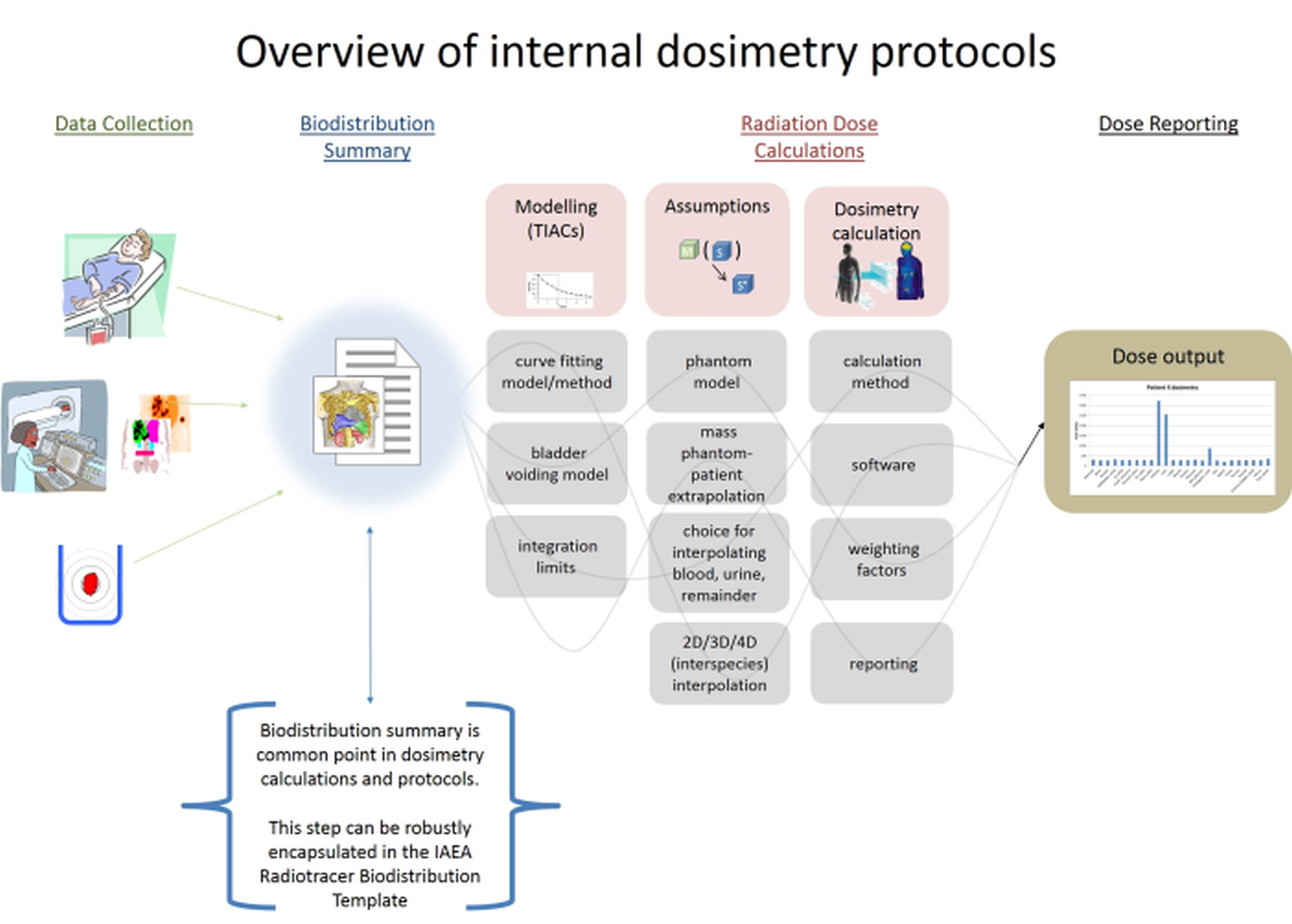
The IAEA Radiotracer Biodistribution Template is organized in information category blocks, as seen in the overview image below.

  • Information about the type of study, technical details, and units for the presented data are defined in SECTION B and SECTION C.
  • Raw data is contained in SECTION D.
  • Time-Integrated Activity Coefficients are contained in SECTION E.
  • Comments clarifying the details of the acquisition/data can be included in SECTION F.


How to use/complete the template

The IAEA Radiotracer Biodistribution Template is meant to document radionuclide biodistribution measurements. This template does not contain dosimetry calculations. However, the intention is that the basic information recorded here may be used for further organ level dosimetry calculations.

To complete the template, enter single field details, then measurement time, volume, activity, and standard deviation, per organ, for the organs that have this information available.

All editable fields initially contain an empty flag (-1). These fields should be changed only when information is available to complete them. It is acceptable/expected that not all fields will be modified. For example, fields delineating organs which are not measured should be left with an empty flag (-1). Measurements only need to be entered for assessed organs/time points.

The template is structured into the following sections:

  • SECTION A - TEMPLATE DESCRIPTION 
    • Instructions for template use
  • SECTION B - ACQUISITION DETAILS 
    • Enter details describing the acquisition of input data
    • Fields include: Subject ID, Species, Sex, Height, Weight, Projected whole body blood volume, Isotope, Radiopharmaceutical, Date of administration, Time of administration, Net injected activity, Software used for image analysis, Institution of acquisition, City of acquisition, Author initials (optional), Inquiry contact email (optional).
  • SECTION C - TECHNICAL DETAILS 
    • Enter details describing technical aspects of data acquisitions
      Main measurement modality, Blood measurement modality, Blood measurement units, VOI/ROI measurement units, Organ VOI/ROI drawing method, Time point units, Volume units, Method of calibration, Attenuation correction applied, Geometric mean applied, Decay correction applied, Scatter correction applied, Partial volume correction applied, Well Counter Calibration Factor, Probe Calibration Factor, SPECT Calibration Factor, Time integrated activity coefficient (residence time)
  • SECTION D1 - MEASUREMENT TIME POINTS 
    • Enter time points associated with the activity measurements used in Section D3
    • For each measurement that will be recorded in section D3, an associated time point needs to be entered
    • Units are specified in section C (suggested hours post injection)
  • SECTION D2 - MEASUREMENT VOLUMES 
    • Enter organ volume, specific to subject studied, for each measurement taken
    • Units are specified in section C
  • SECTION D3 - MEASUREMENT ACTIVITIES 
    • Enter activity measured for each organ/time point studied
    • Units are specified in section C
  • SECTION D4 - MEASUREMENT STANDARD DEVIATIONS 
    • If available, enter the standard deviations associated with the measurements in section D3
  • SECTION E - Time-Integrated Activity Coefficients (TIACs) (residence times) (OPTIONAL) 
    • User may optionally include TIACs calculations derived in section E
    • Time-Integrated Activity Coefficients can be entered with units of hours, or total number of disintegrations, or % of total disintegrations. This detail is specified by user in SECTION C
  • SECTION F – COMMENTS 
    • The comments section has several categories. Please include any comments that would be relevant for an objective reviewer to better understand the study

Examples

Example completed templates can be downloaded for Lu-177 DATATATEI-131.

Types of dose calculation

The IAEA Radiotracer Biodistribution Template supports organ-level dosimetry, which means that activities are assumed to be uniform within organs and the emissions from that organ can be summarized with a single number (i.e. Time-Integrated Activity Coefficients).  Dose calculations are usually calculated from the organ-level biodistribution (recorded in the template) using cross-organ dose factors and the MIRD formalism. The cross-organ dose factors are referred to as “S factors” in the MIRD formalism, or “dose conversion factors” in the OLINDA/EXM software, and are calculated for specific digital phantoms and nuclides and summarize the dose deposited from emissions from one organ to another.

There are alternative methods for calculating internal dosimetry, for example Monte Carlo methods, or dose point kernel methods. Users may explore literature to learn more about these alternative methods.

Advancements in dosimetry methodology based upon use of this template

Many aspects of dosimetry collection would benefit from improved standardization. This template is free for the community and will remain that way. Users who develop tools or publish improvements to dose calculation procedures are encouraged to share their efforts by emailing dosimetry@iaea.org.

Utilization of template for generalized code

The template may be updated in the future, depending on community input. While changes may occur, care will be taken to keep the three letter UID tags (3rd column in sheet) consistently associated with its respective variable. Thus it is recommended to use those tags for building parsing software.

Feedback

We are presenting this template as a resource for the community. We invite feedback on this effort. Please send us comments at dosimetry@iaea.org.

An overview of dosimetry principles can be found on the IAEA human health campus website, in the nuclear medicine dosimetry section.

Acknowledgement

The IAEA Radiotracer Biodistribution Template has been developed by Adam Kesner, PhD, University of Colorado, Denver, CO, USA and Michael Lassmann, PhD, Universitätsklinik Würzburg, Würzburg, Germany with support by Seval Beykan, Universitätsklinik Würzburg, Würzburg, Germany, and Gian Luca Poli, IAEA.

Release of the patient

A patient that has undergone a therapeutic nuclear medicine procedure is a source of radiation that can expose other people in close proximity. External irradiation of others is dependent on the radionuclide used, its emissions, half-life and biokinetics. Excretion (e.g., sweat, saliva, etc) results in the possibility of contamination of the environment and inadvertent exposure to other people.

Important Principles 

While precautions for the public are rarely required after diagnostic nuclear medicine procedures, some therapeutic nuclear medicine procedures, particularly those involving 131I, can result in significant exposure to other people, especially those involved in the direct care and support of patients. Hence, staff, carers and members of the public caring for such patients in hospital or at home require specific consideration.

Patients do not necessarily need to be hospitalized after all radionuclide procedures. Relevant national dose limits must be met and the principle of optimization of protection must be applied. The decision to hospitalize or release a patient should be determined on an individual basis dependent on the residual activity in the patient. Hospitalization will reduce exposure to the public and relatives but will increase exposure to hospital staff. Hospitalization often involves a significant psychological and financial burden that should be analysed and justified. For some patients, hospitalization during and following treatment may be necessary and appropriate. For example, incontinent patients or ostomy patients may require extended hospitalization to ensure safe collection and disposal of biomedical radioactive waste.

The nuclear medicine physician has the responsibility to ensure that a patient, who

has undergone a therapeutic procedure with unsealed sources, is  not discharged from the nuclear medicine facility until properly cleared by the medical physicist. The physicist will verify the activity of radioactive substances in the body is such that the doses that may be received by members of the public, including family members, would meet national criteria and comply with relevant dose limits and dose constraints.

Current recommendations regarding the release of patients after therapy with unsealed radionuclides vary widely around the world. However, the decision to release a patient is based on the assumption that the risk can be controlled when the patient returns to their home. This is generally achieved by combining an appropriate release criterion with well-tailored instructions and information for the patient that will allow them to deal effectively with the potential risk.

General information about nuclear medicine and specific instructions on the management of therapy patients can be found in the Handbook of Nuclear Medicine Physics (IAEA).

General information about nuclear medicine and specific instructions on the management of therapy patients can be found in the Handbook of Nuclear Medicine Physics (IAEA) and in the IAEA Safety Standard (Radiation Protection and Safety of Radiation Sources: International Basic Safety Standards)

Public exposure

Public exposure is exposure incurred by members of the public from radiation sources, excluding any occupational or medical exposure. The sources of exposure of the general public are primarily the same as for workers. Hence, the use of structural shielding and the control of sources, waste and contamination are fundamental to limiting exposure of the public. There are, however, some additional situations that need special consideration. These include the release of patients examined or treated with radiopharmaceuticals.

Important Principles

The unintentional exposure of members of the public in waiting rooms and on public transport is usually not high enough to require special restrictions on nuclear medicine patients, except for those being treated with radioiodine who should receive patient-specific instructions for limiting public exposure. However, the general layout of the nuclear medicine facility should consider the protection of members of the public. The areas for storage and preparation of radiopharmaceuticals must be well separated from public areas such as waiting rooms.

Arrangements should be made to control access of visitors (with added emphasis on controlling access of pregnant visitors or children) to patients undergoing radionuclide therapy and provide adequate information and instructions to these persons before they enter the patient’s room, to ensure appropriate protection. Measurements should be taken in order to restrict public exposure to contamination in areas accessible to the public. Exposure can also occur to those immediately involved with the patient and the general population through environmental pathways including sewerage and discharges to water. For diagnostic patients, there is no need for the collection of excreta and ordinary toilets can be used. For therapy patients, there are various policies in different countries, but, in principle, the clearance criteria should follow a dilution and decay methodology. Eventually, much of the activity initially administered is discharged to sewers. Storing a patient’s urine after therapy appears to have minimal benefit as radionuclides released into modern sewage systems are likely to result in doses to sewer workers and the public that are well below public dose limits. Once a patient has been released from hospital, the excreted radioactivity levels are low enough to be discharged through the toilet in their home without exceeding public dose limits. The guidelines given to patients will protect their family, carers and neighbours, provided the patient follows these guidelines.

The IAEA book on Nuclear Medicine Physics provides a detailed and well-structured overview to the topic of Nuclear Medicine.

Stay in touch

Newsletter