The German physicist Wilhelm Röntgen was the first person to discover the potential use of X-rays for imaging in 1895, when he created the radiograph, an image produced by X-rays instead of light. The first X-ray photograph, revealing the bones of his wife’s hand, took the world by storm, revolutionising medicine and earning him the first Nobel Prize in physics. Today, diagnostic radiology has become one of the most powerful tools in modern medicine. Radiology is essential for the timely and accurate diagnosis of a range of conditions from broken bones to cancer, enabling effective treatment to begin as early as possible.
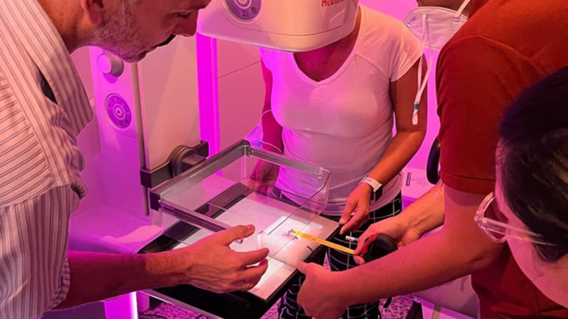
As vital radiology equipment applies ionizing radiation to patients, its performance needs to be monitored to ensure safe and effective use. The IAEA has recently released a new Handbook of Basic Quality Control Tests for Diagnostic Radiology as a quick reference guide on how to perform quality control tests and draws attention to the common issues and mistakes that could undermine the results or the evaluation of a given test.
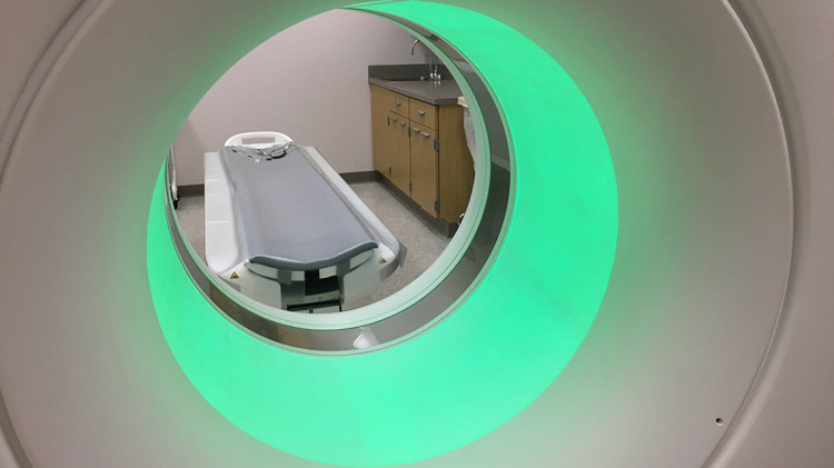
Medical imaging technology has evolved remarkably over the past century, shifting steadily from analogue to digital radiology, from first generation computed tomography (CT) to hybrid sophisticated systems. These technologies involve the use of X-rays, a form of ionizing radiation, passing through a section of a patient’s body. Ionizing radiation, when used safely and securely by trained professionals for medical diagnosis, is safe. The benefits of medical diagnostic imaging far outweigh any risks. However, it is essential that equipment used in radiology treatment is subject to strict quality controls, as exposure to high levels of ionizing radiation can lead to cellular damage in humans.
Monitoring the performance of radiology equipment is done via a process to ensure safe and effective use. This process starts from basic quality control (QC) and builds up to comprehensive quality management systems, where every aspect of patient care is integrated.
However, in many countries, a significant number of X-ray machines and equipment used in diagnostic radiology are not under a regular quality assurance programme, mainly due to a lack of trained professionals and corresponding guidance. The IAEA developed the QC handbook that compiles all existing performance tests at a global level to fill this gap.
"Imaging equipment is often left without proper performance supervision for long periods of time, only taking place during inspection or licencing process,” said Harry Delis, a diagnostic radiology medical physicist at the University of Patras, Greece, and a contributor to the publication. “Such a guidance document will facilitate establishing or strengthening QC programmes of radiology departments around the world.”
The Handbook is divided into sections dedicated to a specific imaging modality, such as radiography, fluoroscopy, CT and mammography. The publication was reviewed by 30 international experts and endorsed by three scientific organizations, highlighting its importance and reliability for professionals in the field. These include: the American Association of Physicists in Medicine (AAPM), the European Federation of Organisations for Medical Physics (EFOMP) and the International Society of Radiographers and Radiological Technologists (ISRRT).
“The handbook, supported through scientific and professional collaboration, is a useful tool for regular quality assurance implementation by diagnostic imaging medical physicists,” said May Abdel-Wahab, Director of the IAEA Division of Human Health. “The IAEA promotes a comprehensive approach to quality through quality management systems and supports the development of guidance and training of appropriate staff to maintain a high level of quality.”
The publication is freely available here.