Presenter: PhD Ruben Pauwels Date of broadcast: 23 January 2018, 3 pm CET
About the webinar
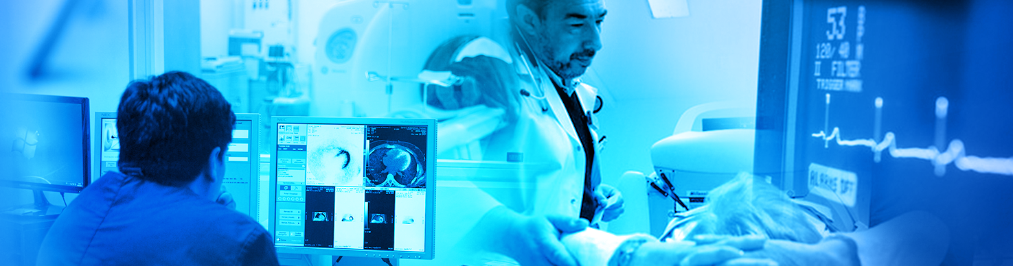
Although cone-beam computed tomography (CBCT) has shown a high diagnostic efficacy for several dental and maxillofacial applications (e.g. implant planning), its use has led to an increased collective patient dose in dental practice. Therefore, it is essential to optimize exposures to the lowest achievable level.
Dental CBCT in particular has shown a considerable leeway for optimization, with doses between an optimized and non-optimized scan possibly varying by an order of magnitude or more.
In this presentation, an overview of different practical approaches towards optimization of dental CBCT exposures will be provided. This process should involve different stakeholders, including the end-user (e.g. oral radiologist, dental practitioner, radiation technologist), medical physicist and manufacturer. It requires a considerable degree of general knowledge regarding the effect of different exposure parameters (e.g. field of view, tube voltage, tube current, exposure time), but at the same time it requires an individualized approach due to the variety of CBCT models found on the market as well as the highly different imaging requirements for specific clinical applications in dentistry.
In the final part of the presentation, a few misconceptions regarding dental CBCT imaging that may affect the optimization process will be addressed: effect of exposure settings on metal and motion artefacts, efficacy of patient shielding, and (lack of) applicability of Hounsfield Units.
Learning objectives
To become familiar with the general principle of CBCT image acquisition.
To understand the effect of each exposure parameter on image quality and patient dose in CBCT.
To be able to optimize CBCT exposures at the level of individual patients (for end-users).
To be able to guide the end-user in the optimization process (for medical physicists and other stakeholders).
About the presenter
Ruben Pauwels obtained MSc degrees in Biomedical Sciences and Medical Imaging, as well as a PhD degree in Biomedical Sciences at the Catholic University of Leuven (KU Leuven), Belgium. He is currently active as as researcher at the OMFS-IMPATH research group, KU Leuven and as a research fellow at Chulalongkorn University, Bangkok, Thailand. Since 2006, his research has primarily focused on dental cone-beam computed tomography, covering an array of topics such as clinical and technical image quality assessment, radiation dosimetry, optimization of exposures, characterization of bone quantity and quality, and artefact reduction.