
Training material
The training material on this webpage is available for download.
The purpose of the material is to train the trainers, who are hospital personnel e.g. medical physicists/radiation protection officers, medical and paramedical staff concerned in the corresponding specialty.
Approved training packages may be copied, distributed, displayed, incorporated in customized presentations and used for non-commercial use as long as the source of the material is referenced to the IAEA approved training package.
It has been developed in collaboration with World Health Organization (WHO), Pan American Health Organization (PAHO), International Labour Organization (ILO), International Society of Radiology (ISR), International Organization for Medical Physics (IOMP), International Society of Radiographers and Radiological Technologists (ISRRT), FDI World Dental Federation, International Association of Dento-Maxillofacial Radiology, and Image Gently Alliance.
Training material
Diagnostic and interventional radiology →
Digital radiology →
Paediatric radiology →
Radiation dose management in computed tomography (CT) →
Radiotherapy →
Radiotherapy: Prevention of accidental exposure →
Safety and quality in radiotherapy →
Nuclear medicine →
Cardiology →
PET/CT →
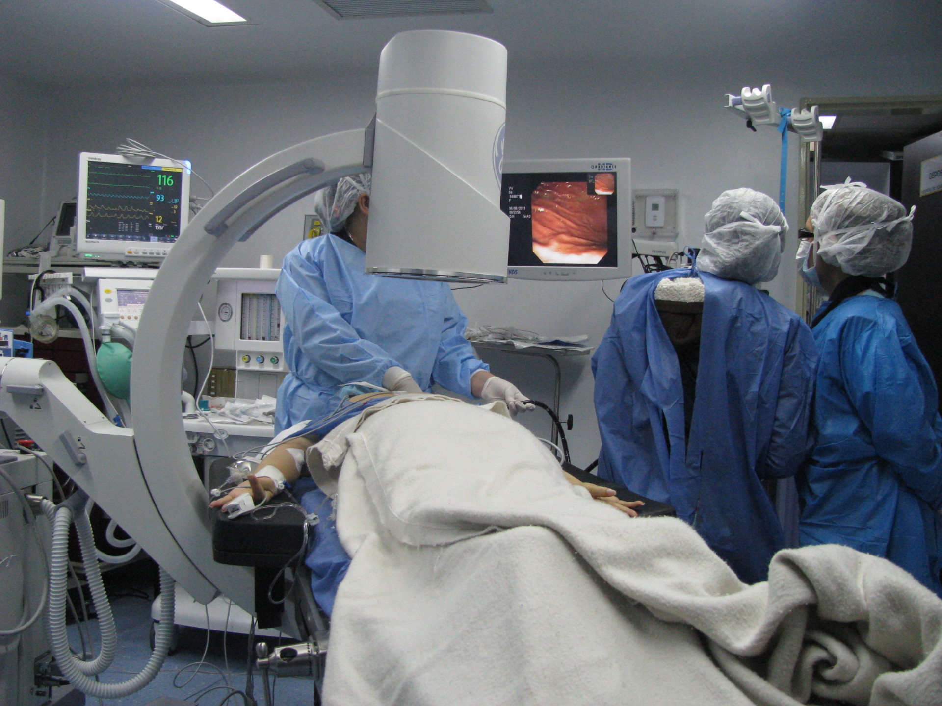
Doctors using fluoroscopy outside radiology (Urologists, Gastroenterologists, Orthopaedic surgeons etc.) →
Dental radiology →
Radiation Safety Culture Trait Talks Handbook →
Radiation Protection in Interventional Procedures: Practical Tutorials →
Diagnostic and interventional radiology
Lectures:
- 00. Principles of radiation protection and motivation for the course
- 01. Overview of radiation protection in diagnostic radiology
- 02. Radiation units and dose quantities
- 03. Biological effects
- 04. International system of radiation protection
- 05. Interaction of radiation with matter
- 06. X-ray production
- 07. X-ray beam
- 08. Factors affecting image quality
- 09. Medical exposure BSS
- 10. Patient dose assessment
- 11. Quality assurance
- 12. Shielding and X-ray facility design
- 13. Occupational exposure: Part 1-2
- 14. Radiation exposure in pregnancy
- 15. Optimization of protection in radiography: Part 1-2
- 16. Optimization of protection in fluoroscopy: Part 1-2
- 17. Optimization of protection in interventional radiology: Part 1-2
- 18. Optimization of protection in CT
- 19. Optimization of protection in mammography
- 20. Optimization of protection in digital radiology
- 21. Optimization of protection in paediatrics
- 22. Optimization of protection in dental radiology
- 23. Organizing a QA program in diagnostic radiology
Practical exercises:
- 12. Shielding and X-ray facility design: Part 1-2
- 15. Optimization of protection in radiography: Part 1-4
- 15. Optimization of protection in radiography: Part 5-6
- 15. Optimization of protection in radiography: Part 7-9
- 16. Optimization of protection in fluoroscopy: Part 1-6
- 18. Optimization of protection in CT
- 19. Optimization of protection in mammography: Part 1-10

Lectures:
- 01. Fundamentals of Digital Radiography
- 02. Exposure indicators and patient dose estimation in CR and DR
- 03. Optimization in CR and DR
- 04. Optimisation of Digital Fluoroscopy
- 05. Digital Radiographic Image Processing
- 06. Avoiding Artefacts in Computed Radiography
- 07. Avoiding Artefacts in Digital Radiography
- 08. Optimising DR Displays
- 09. Picture Archival and Communication System (PACS)
- 10. Practical Exercises
- Clinical Problems Oriented Flowcharts
- Topics & Objectives

Lectures:
- 01. Why Talk About Radiation Protection during Radiological Procedures in Children
- 02. Understanding Radiation Units
- 03. Radiation Protection of Children in Screen Film Radiography
- 04. Radiation Protection of Children in Digital Radiography
- 05. Radiation Protection of Children in Fluoroscopy
- 06. Radiation Protection of Children During Computed Tomography
- 07. Radiation Protection of Children in Interventional Radiology and Cardiology
- 08. Standards and Guidelines in Radiological Procedures in Children
- 09. Quality Assurance in Paediatric Radiological Procedures
- 10. Organization of a Paediatric Radiology Department
Lectures:
- 01. Appropriateness and justification for CT
- 02. Overview of scan parameters
- 03. Tube current and AEC
- 04. Tube potential in CT
- 05. Detector configuration, pitch and speed
- 06. CT dose metrics and tracking
- 07. Chest CT key aspects
- 08. Key aspects of abdomen CT
- 09. Key aspects of head CT
- 10. Key aspects for pediatric CT
- 11. Strategies for CT in pregnant patients

Lectures:
- 00. General introduction
- 01. The aim and role of radiotherapy
- 02. Radiation physics: Part 1-2
- 03. Biological effects of ionising radiation: Part 1-2
- 04. Principles of radiation protection and the international safety standards: Part 1-2
- 05. Properties and safety of radiotherapy sources and equipment (external beam therapy): Part 1-4
- 06. Properties and safety of radiotherapy sources and equipment (brachytherapy): Part 1-3
- 07. Design of facilities and shielding calculation: Part 1-2
- 08. Occupational exposure
- 09. Medical exposure
- 10. Medical exposure: optimization in external beam therapy: Part 1-8
- 11. Medical exposure: optimization in brachytherapy: Part 1-3
- 12. Medical exposure: Quality assurance
- 13. Medical exposure: Potential and accidental exposures: Part 1-2
- 14. Transport safety
- 15. Security of sources, and disposal of disused sources
- 16. Discharge of patients
- 17. Protection of the public
- 18. Organization and implementation of a radiation protection programme
Practical exercises
- 01. The aim and role of radiotherapy
- 02. Radiation Physics: Part 1-2
- 03. Biological effects of ionising radiation: Part 1-2
- 05. Properties and safety of radiotherapy sources and equipment
- 07. Design of facilities and shielding calculation
- 10. Medical exposure: Optimization of protection in external beam therapy Part 1-3
- 11. Medical exposure: Optimization of protection in brachytherapy
- 12. Medical exposure: Quality assurance Part 1-2
- 18. Organization and implementation of a radiation protection programme

Lectures:
- 0.1 Introduction
- 1.1 BSS general requirements
- 1.2 BSS Section 3 - medical exposure
- 2.1 Incorrect decay data - USA
- 2.2 Erroneous use of TPS - UK
- 2.3 Accelerator software problems - USA & Canada
- 2.4 Computer file not updated - USA
- 2.5 Incorrect repair of accelerator - Spain
- 2.6 Miscalibration of beam - Costa Rica
- 2.7 Error in TPS data entry - Panama
- 2.8 Accelerator interlock failure - Poland
- 2.9 HDR unit malfunction - USA
- 2.10 Accident update, some newer events - UK, USA & France
- 3.1 Other cases - external beam therapy
- 3.2 Other cases - brachytherapy
- 3.3 Incidents in any clinic
- 4.1 Clinical consequences of accidental exposures in radiotheraphy
- 5.1 Preventive measures
- 5.2 Reporting and investigating
- 6.1 Source not under control - Brazil
- 6.2 Source not under control - Mexico
- 6.3 Source not under control - Turkey & Thailand
Lectures:
- 01. Introduction
- 02. Major Incidents in Radiotherapy
- 03. Learning From Incidents
- 04. Process Maps, Severity Metrics, Basic Causes & Safety Barriers
- 05. Reporting Incidents Using Safron
- 06. Root Cause Analysis 1. Human Factors & Basic Causes
- 07. Root Cause Analysis 2. Safety Barriers & Preventive Actions
- 08. Failure Modes and Effects Analysis
- 09. Fault Tree Analysis
- 10. Safety Culture
- 11. Useful Resources
- 12. And Now What? Enhancing Quality and Safety in Your Clinic
Lectures:
- 00. Introduction to nuclear medicine
- 01. Biological effects
- 02. Radiation physics
- 03. Principles of radiation protection
- 04. Safety of sources and design of facilities
- 05. Occupational protection
- 06. Medical exposure
- 07. Optimization of medical exposure: Diagnosis
- 08. Optimization of medical exposure: Therapy
- 09. Quality assurance
- 10. Radioactive waste
- 11. Potential exposure
- 12. Protection of the general public
- 13. Organization of radiation protection in nuclear medicine
Notes:
- 01. Biological effects
- 02. Radiation physics
- 03. Principles of radiation protection
- 04. Safety of sources and design of facilities
- 05. Occupational protection
- 06. Medical exposure
- 07. Optimization of medical exposure: Diagnosis
- 08. Optimization of medical exposure: Therapy
- 09. Quality assurance
- 10. Radioactive waste
- 11. Potential exposure
- 12. Protection of the general public
- 13. Organization of radiation protection in nuclear medicine
Practical excercises:
- 01. Pulse height analysis
- 02. Simulated inspection of a nuclear medicine facility
- 03. Workplace monitoring
- 04. Decontamination
- 05. Shielding of sources
- 06. Quality control of activity meter
- 07. Quality control of gammameter
- 08. Gammacamera imaging

Lectures:
01. Why talk about radiation protection in cardiology?
02. Talking about radiation dose
03. What radiation effects are possible? (besides skin injuries)
04. X ray production and angiography equipment
05. Patient dose management: Part 1-2
06. Standards and guidance
07. Occupational exposure and protective devices
08. Image quality in cardiac angiography
09. Optimization of radiation protection in cardiology
10. Radiation protection in paedriatic interventional cardiology
11. Cardiac CT - radiation doses, dose management and practical issues
12. Examples of Good & Bad Practice (physical factors): Part 1-2
Lectures:
- 01. Introduction to Radiation Protection in PET/CT
- 02. PET/CT Technology
- 03. Medical Exposure - BSS Requirements
- 04. Protection issues in clinical methodology
- 05. Facility design
- 06. Protective equipment
- 07. Personal & work place monitoring
- 08. Staff and public doses
- 09. Transport safety, source security & dealing with waste
- 10. Written procedures and organization
- 11. Quality control
- 12. SPECT/CT technology & facility design
Lectures:
- 01. Overview of radiation protection
- 02. Understanding radiation units
- 03. What can radiation do?
- 04. Anatomy of fluoroscopy & CT Fluoroscopy Equipment
- 05. How do I reduce my radiation risk?
- 06A. Radiation protection for patients in orthopaedic surgery
- 06B. Radiation Exposure in Gastroenterology
- 06C. Other medical specialties that use fluoroscopy
- 07. International standards and recommendations

Lectures:
- 01. General Principles of Radiation Protection
- 02. Special Considerations for Radiation Protection in Children
- 03. X-ray Production and Interaction: Image Formation and Image Quality
- 04. General Principles of Film and Digital Radiography
- 05. Fundamentals of Intraoral Radiography
- 06. Fundamentals of Panoramic Radiograhy
- 07. Fundamentals of Extraoral Projectional Radiography
- 08. Fundamentals of CT and CBCT
- 09. Justification and Appropriate Use of Dental Radiology
- 10. Quality Assurance in Dental Radiology
- 11. Optimization of Protection of Patients in Dental Radiology
- 12. Protection of Workers and Public in Dental Radiology
- 01. Individual Responsibility
- 02. Questioning Attitude
- 03. Effective Safety Communication
- 04. Leadership Responsibility
- 05. Decision-making
- 06. Respectful Work Environment
- 07. Continuous Learning
- 08. Problem Identification and Resolution
- 09. Environment for Raising Concerns
- 10. Work Processes
Radiation Protection in Interventional Procedures: Practical Tutorials

The tutorials will download as zipped folders. To unzip all the contents of the zipped folder, press and hold (or right-click) the folder, select Extract All. Please do this in the zipped folder, which you will find in your downloads. Once the files are extracted, to view the interactive video please click on story.html.
- 01. Effect of fluoroscopic projection on staff radiation dose (English) (To download click here)
- 02. Effect of collimation on staff radiation dose (English) ( To download click here)
- 03. Virtual collimation and its effect on staff radiation dose (English) (To download click here)
- 04. Effect of patient size on staff radiation dose (English) (To download click here)
- 05. Effect of ceiling suspended screens on staff radiation dose (English) (To download click here)
- 06. Effect of lead aprons on staff radiation dose (English) (To download click here)
- 07. Effect of fluoroscopy mode on patient and staff radiation dose (English) (To download click here)
- 08. Effect of magnification on patient and staff radiation dose (English) (To download click here)
- 09. Effect of patient–X ray tube distance on patient radiation dose (English) (To download click here)
- 10. Effect of patient–image receptor distance on staff radiation dose (English) (To download click here)
- 11. Effect of radiation protection gloves on patient and staff radiation dose (English) (To download click here)
- 12. Real time dose monitoring systems (English) (To download click here)
- 13. Radiation and cataract (English) (To download click here)