You are here

A Window Inside the Body and COVID-19

Medical imaging during the global pandemic

Nicole Jawerth

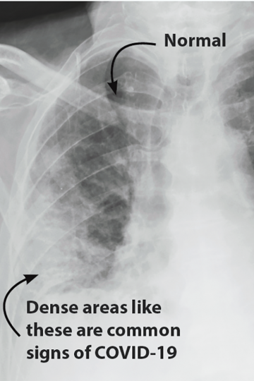
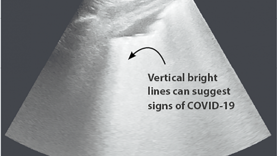
Getting images of what’s happening inside of people’s bodies is helping health professionals to evaluate and better understand the COVID-19 disease, which is caused by a newly discovered coronavirus.

“Diagnostic imaging is a window into the body,” said Olivier Pellet, a radiologist at the IAEA. “It has allowed us to uncover complications, such as lesions, pneumonia or blood clots in the lungs. Every day we learn about the virus and its impact on the human body, while discovering new COVID-19-related signs and symptoms we have not seen before, even when a person may otherwise appear asymptomatic.”

Every day we learn about the virus and its impact on the human body, while discovering new COVID-19-related signs and symptoms we have not seen before, even when a person may otherwise appear asymptomatic.
Olivier Pellet, radiologist, IAEA

Medical imaging has been used worldwide for over a hundred years to diagnose, monitor and assist in the treatment of many health conditions, such as cancer, infectious diseases, heart disease and neurological disorders. Many countries have been working with the IAEA for decades to build up and maintain their radiation medicine services, including diagnostic imaging.

While there are a range of imaging techniques available, the three most widely used methods for evaluating COVID-19 patients are chest X-rays, chest computed tomography (CT) and lung ultrasound.

“These three techniques are complementary and offer options for evaluating how COVID-19 affects different organs at different stages,” said Pellet. “The reason they are used on the lung and chest area is because respiratory symptoms have been known to be among the first signs of COVID-19.”

Although diagnosing COVID-19 relies on identifying the virus using laboratory tests such as reverse transcription–polymerase chain reaction, medical imaging is widely used to evaluate patients at various stages of the disease, particularly for those with moderate, severe or critical cases.

Refining and adapting

Since the global spread of COVID-19 began in early 2020, health professionals have had to adapt and improve how they use these techniques to ensure that they are justified, appropriate and safe for diagnosing the disease.

“Radiologists and other imaging specialists have had to figure out which procedures to use for COVID-19 and when, how to accurately analyze and identify the disease’s symptoms in medical images, and how to adapt their work to protect staff and patients from infection while continuing to provide other essential and critical health services,” Pellet said.

They have also had to stay vigilant about striking the right balance: using too little radiation makes the images unclear, but too much puts the patient at risk of receiving an unnecessary dose of radiation. Similarly, taking more scans than needed means unnecessary exposure, while too few could mean missing out on critical information to help a patient.

“Any time radiation is used in medicine, it needs to be justified and optimized to ensure that the procedures are effective, while also keeping patients and workers safe,” said Miroslav Pinak, Head of the Radiation Safety and Monitoring Section at the IAEA. “During a pandemic situation, when normal procedures and workflows are interrupted, attention must be paid to continuing to maintain high standards of radiation protection while also integrating measures required to minimize the spread and impact of COVID-19.”

To support these efforts, the IAEA has provided health professionals with a wide range of resources, such as webinars, articles and technical guidance documents, related to COVID-19 and radiology, nuclear medicine and radiation protection.

Chest X-rays

Chest X-ray of a patient with COVID-19 pneumonia. Both lungs are showing signs, particularly the right. (Photo: L. Zanoni/Nuclear Medicine Division, Bologna University Hospital Authority St. Orsola-Malpighi Polyclinic)


X-rays are a type of radiation. Many people are most familiar with them for their use in diagnosing a broken bone or examining teeth.

Health care professionals can harness X-ray radiation using an X-ray machine. When a patient is placed in the machine, a carefully selected dose of X-rays passes through the targeted area of the body. The thicker and denser parts of the body, such as bone, let fewer X-rays through, while softer, thinner parts let more through. As the X-rays come out on the other side of the body, a specialized detector captures their pattern. This creates an image of the structures inside the body and their changes.

For evaluating COVID-19, X-rays are taken of a person’s chest to get a look at the lung tissue. This is used for patients with respiratory symptoms of COVID-19. X-rays are also used for monitoring the evolution of the disease and making treatment and follow-up decisions, such as whether to admit a patient to the hospital or send a patient with severe symptoms for a CT scan.

“Given that X-ray machines are often readily available in care centres, many health care professionals already have access to these tools to help their countries deal with COVID-19,” Pellet said. “Some X-ray machines are also portable, lightweight, and easy to handle and decontaminate, which is critical in a pandemic, particularly in medical triage areas or makeshift hospital environments.”

Chest CT

Chest CT of a patient with COVID-19 pneumonia. Both lungs are affected, particularly the right. (Photo: L. Zanoni/Nuclear Medicine Division, Bologna University Hospital Authority St. Orsola-Malpighi Polyclinic)

A computed tomography (CT) scan is a set of multiple X-ray images. A CT machine rotates around the patient and rapidly sends X-rays through the body from multiple angles. A ring of hundreds of specialized detectors around the body track the pattern of the X-rays. This is then processed by the machine’s powerful computer to create detailed images constructed out of very thin slices of the body, up to 0.3 mm wide, often in 3D. For a CT scan of the chest, which is the area of the body typically scanned when evaluating COVID-19, hundreds of images are generated to cover the whole chest area.

CT machines are more sophisticated, more expensive and not as widely available as X-ray machines. They are also harder to decontaminate, which can take more than 20 minutes, said Pellet. “CT scans provide highly detailed information and to do that they use more radiation than an X-ray machine. They should therefore only be used when appropriate for the patient’s case.”

Ultrasound

Lung ultrasound of a patient with COVID-19 pneumonia. (Photo: C. Serra/Nuclear Medicine Division, Bologna University Hospital Authority St. Orsola-Malpighi Polyclinic)


Ultrasound machines use high-frequency soundwaves instead of radiation to create an image. A probe connected to an ultrasound machine sends and receives millions of soundwaves every second through the target area of the body, which, in the case of COVID-19 patients, is typically the lungs. When the waves hit a boundary, such as between soft tissue and fluid or soft tissue and bone, they echo back to the probe. The probe tracks the distance and intensities of the echoes and translates this into images.

Ultrasound machines are low cost and more widely available than X-ray and CT machines. Being small, portable and easy to decontaminate, they can be easily used at patient bedsides, in an ambulance or in a triage situation. Since radiation is not involved, they can also be used more often without posing additional risks to patients and health care workers.

As ultrasound images are dynamically and instantly displayed on a screen, a trained health care professional can evaluate a patient on the spot. Ultrasound imaging of the lungs is a good starting point for evaluating patients displaying respiratory symptoms that could be signs of COVID-19, as it can display images that are strongly suggestive of the disease. However, as lung ultrasound only explores the periphery of the lungs and is user dependent so concrete and detailed images provided through chest X-rays and chest CT scans are needed for a conclusive diagnosis of COVID-19, as well as to follow and monitor the evolution of the disease in the patient.


June, 2020
Vol. 61-2

Stay in touch

Newsletter