
Many people have experienced a scan in the nuclear medicine department of a hospital or clinic. It may seem a straightforward procedure for the patient, but the technology involved is a complex interaction of nuclear medicine and radiology imaging techniques that has evolved over 50 years into sophisticated hybrid systems known as SPECT/CT, or Single Photon Emission Computed Tomography/Computed Tomography.
For the safe and effective use of this technology a rigorous quality control regime is necessary, which is the theme of a recent IAEA publication SPECT/CT Atlas of Quality Control and Image Artefacts. The Atlas is intended as a guide for professionals in nuclear medicine: medical physicists, nuclear medicine specialists and medical radiation technologists.
“Although this technology provides a wealth of diagnostic information, it is important that practitioners understand the principles of image formation and are fully aware of the potential pitfalls and image artefacts that can be encountered in clinical practice,” said Debbie van der Merwe, Head of the Dosimetry and Radiation Physics Section at the IAEA. “This newly published Atlas presents an overview of these potential pitfalls as well as the quality control procedures and standards required in SPECT/CT.”
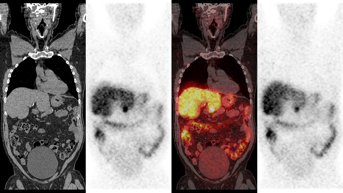
The Atlas is presented in three main sections, firstly reviewing the usage of CT and SPECT images and the various systems currently available. Secondly, the quality control procedures for the operation of SPECT and SPECT/CT systems in routine clinical practice are outlined. The concluding section presents a broad selection of 39 well-illustrated case studies of potential image artefacts from different sources, ranging from hardware malfunctions to user- and patient-induced artefacts. In addition, descriptions are given of their causes and the techniques used to identify them and avoid their recurrence.
“A useful and reliable diagnosis from SPECT/CT imaging depends on the accurate interpretation of the images,” said Ms. van der Merwe. “Possible image abnormalities related to machine, user or patient factors might not be recognized by nuclear medicine professionals, which could ultimately hinder optimal patient management.” She continued, “the case studies illustrated in the SPECT/CT Atlas will help practitioners identify and eliminate these abnormalities.”
The SPECT/CT Atlas of Quality Control and Image Artefacts is published as part of the IAEA’s Human Health Series. Publications in this series present analyses and provide information of an advisory nature, including guidelines, codes and standards of practice, and quality assurance materials.


