To stop diseases like cancer, doctors need to see what is happening inside the body. One nuclear technique that does this is Positron Emission Tomography (PET), a non-invasive technique that allows doctors to track organ function at a molecular level, therefore revealing intricate health changes earlier than if other diagnostic techniques were used.
Positron Emission Tomography, also called PET imaging or a PET scan, uses a technology that is completely different from either the CT Scan (which uses X-Rays), or the MRI. It images the body's soft tissues, sending back pictures that tell the physician how certain clusters of cells are acting.
To conduct a PET scan, the patient is injected with a sugar analogue labelled with a radioisotope, able to identify tumour cell deposits. "The amount of radioactivity is very low. So the exposure is less than what you would get from a CT Scan," says Homer Macapinlac, Chairman of the Department of Nuclear Medicine at the MD Anderson Medical Center in the US.
"We inject it intravenously and it distributes exactly like sugar would in the body. But the amount is very small. It's called a trace amount. So it does not affect the patient's body," he says.
PET scans are used quite often in identifying the size, shape and location of cancers. "Since cancer cells use more sugar than normal cells and malignant cancers in general use more glucose than benign ones, PET scans are extremely useful. For example, it can save patients from having to undergo biopsies or unnecessary surgeries; because a PET scan, which is performed on the whole body, can accurately show what is happening inside the body," says Macapinlac.
This technology is quite expensive, and as such is most widely used in developed countries. Some developing countries are also adopting the technology. Between 30% and 35% of IAEA Member States have at least one PET centre or are in the advanced stages of planning a new PET centre.
The Right Place
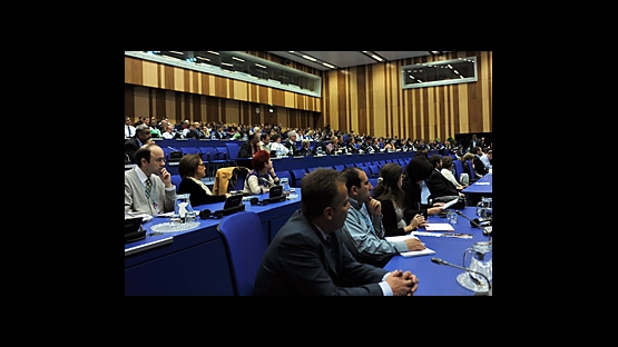
The IAEA's mandate includes promoting the application of nuclear science in all fields, including human health, especially among those countries that have yet to benefit from these applications. As a result, the IAEA organised the International Conference on Clinical Positron Emission Tomography (PET) and Molecular Nuclear Medicine, which Maurizio Dondi, Head of the IAEA Nuclear Medicine Section, describes as "a good way for experienced countries and countries new to the technology's application to teach and learn respectively".
"Our colleagues in developing countries can hardly afford to go to the major international conferences, which are prohibitively expensive. That's why we've organised our own conference, bringing the major experts in the field together with colleagues from developing countries, who can then be exposed to the latest advances in PET technology," says Dondi.
The Agency subsidised the costs for experts and participants from developing Member States.
The Right People
The IAEA's role is to help Member States implement nuclear technology safely and efficiently, which includes training those who are going to be using it - doctors, medical physicists, radiopharmacists and radiochemists. As such the Agency supports various national and regional projects worldwide to build the necessary human resource capacity, and provides documents, guidance, and standards for quality of practice.
"We hope that when they go back to their institutions they will apply more advanced applications with greater standards of quality in order to provide better service to cancer patients," says Dondi.


